
| Quarterly New Technology Update | April 2, 2014 |
In This Edition
- Top Story
- Product Review: Santinelli
- Product Review: Diopsys
- New Technology: Apex EDI
- New Technology: ClearPath
- New Technology: Sonomed Escalon
- Give Us Your Feedback
Tech Update
The Gazette
Archives
The Exchange Booth Quick Reference Guide
- Apex EDI: #523
- ClearPath: #534
- Diopsys: #927
- Santinelli: #1039
- Sonomed Escalon: #315


Contact Vision Source® at 888-558-2020 or [email protected]
Top Story
Welcome to the Quarterly New Technology Update
It is amazing the number of new technologies that we are able to see, as we travel to the various meetings across the country. Most of these companies with new technologies are eager to become vendor partners and offer these products and technologies to Vision Source® members. As a new service to members, we will provide a quarterly update to members on the offerings from existing and new vendor partners with new technologies.
These products and services will be evaluated in a clinical setting, and we will try and point out the applications and advantages to these technologies so that you can gauge for yourself whether it is the right fit for your practice.
 |
 |
John A. McCall, OD |
Carl Spear, OD, FAAO |
Product Review: Santinelli
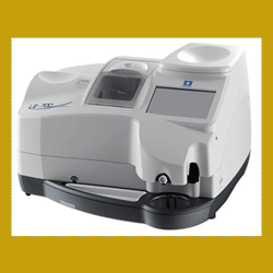
Dr. Spear Tests the Santinelli LE- 700 Edger
| Booth #1039 |
In August of 1998, I bought my first edger for in-office finishing, and it was one of the best decisions I ever made for the practice. We recently had the opportunity to do an in-office evaluation of Santinelli's new LE-700 edger.
We first noticed the compact footprint and integrated blocker. Unlike my first edger 16 years ago that took up an entire room and required two people to lift, the LE-700 only weighs 70 pounds and is ideal unit for offices with limited space. The LE-700 features tracer-free technology, which measures the demo lenses and patterns within the processing chamber. In addition to tracing the demo lens circumference, the front curve of the lens is measured to obtain 3-D tracing data and perform 3-D edging. This 3-D visualization allows you to see how the lens will look and make any adjustments prior to cutting the lens.
We had the edger out of the box and set up to run in less than an hour. As we began to use the unit, the next-step wizard technology guided us through the entire tracing, blocking, edging and grooving process using an intuitive touch-screen display and quick-read icons. If you have never edged before, you'll definitely appreciate the step-by-step process that eliminates the need to have an experienced lab person. In addition, the training and support provided by Santinelli has been excellent so you can get answers to any questions you may have quickly and easily.
Add-ons for the edger include a front-loading frame tracer, which gives you the capability to handle high-wrap frames, as well. We tried this feature and found it extremely reliable, and it allowed us to edge several jobs we would have never been able to do with our previous system.
If you are considering adding an edger to your practice (and if you don't yet have an edger, you should be considering doing this) the Santinelli LE-700 would be a great addition. It is compact, lightweight, easy to operate and surprisingly quiet, and the pricing makes it affordable and an excellent value.
For more information contact them at 800-644-3343 and go see Santinelli's LE-700 and more at its Vision Source® Exchange booth (#1039).
Product Review: Diopsys
Dr. McCall Reviews the Diopsys® NOVA Vision Testing System
| Booth #927 |
I've had this system in my office for almost two-and-a-half years, and I can honestly say it is one of the best investments that I've made while practicing optometry. It gives me objective data on the function of the vision system that I couldn't get any other way, and it pays me well for its use.
I started with the Diopsys® NOVA-VEP in 2011, primarily for the early detection benefits that visual evoked potential provides in glaucoma cases. I quickly learned that the device is also extremely helpful in helping to detect optic neuritis, and I've had several patients diagnosed with MS after a neurology referral. One of the more interesting uses is for traumatic brain injury (TBI). I can assess the impact a TBI has had on my patient's vision system, and easily track if the patient is getting better or worse at subsequent visits.
Last year I purchased the Diopsys® NOVA-ERG module, which increased our ability to diagnose and track maculopathies and plaquenil toxicities, and again our revenue increased dramatically. The pattern ERG tests the function of the retina, whereas the VEP tests the function of the entire visual pathway. We've been using the ERG mostly for our established glaucoma patients. We're in good company on this, as the Bascom Palmer Eye Institute recently published a study that showed changes in pattern ERG results can be detected approximately eight years sooner than changes in RNFL thickness.
 |
The device makes sense financially, too. These tests receive the highest testing reimbursement in my office. The American Medical Association reports that 2014 National Payment Allowables for the VEP CPT Code – 95930 – range from $96.27 to $179.71. The allowables for the ERG CPT Code – 92275 – range from $121.45 to $208.14. VEP is not part of the Multiple Procedure Payment Reductions, and even though ERG is, it's almost always the highest-reimbursed test during the patient's visit, so you get paid the full amount. You can also do the VEP and ERG with other tests during the same visit. I was able to pay off my device within months.
Diopsys provides an outstanding training program, and has supported me and other Vision Source® members in an outstanding fashion. The company really understands how an optometric practice works, and offers its training around how we run our practices. We have our own Clinical Application Specialist who came in initially for a two-day training and is always available to answer questions. Diopsys also has an Educational Review Program, where its neuro-ophthalmologist on staff will review case files with you so you understand how the VEP or ERG results fit into your overall workup of the patient. The company has videos, webinars, case studies and more to help both the doctors and staff understand theitechnology.
I highly recommend visiting the Diopsys booth (#927) at this year's national meeting. You will not be disappointed.
New Technology: Apex EDI
Apex EDI Helps You Locate More Revenue!
Prepare for 2014 insurance changes with new payment technologies
| Booth #523 |
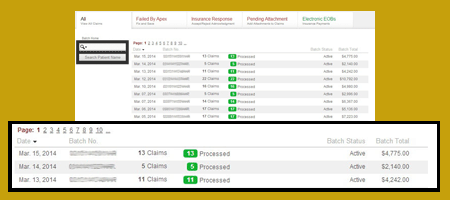
The Vision Source® claims portal, (Powered by Apex EDI) – now includes Apex EDI Revenue Locator, which allows you to match EOB, paymenst and missing revenues efficiently—saving you time and boosting your bottom line.
Many sources promote revenue cycle management, but none are as complete as Apex EDI. Submitting claim reimbursements through the complete revenue cycle keeps you updated through all steps of claim processing. After the claim completes processing, you receive an EOB that Apex evaluates to provide you complete payment information. Here are the benefits.
- Revenue Locator—Presents the claim history, shows your initial claim charge, the process amount, the paid amount, the check number and the amount still owed to you after the claim is processed. Immediately you see where your money is.
- Revenue Difference—Details the difference paid in your claim. The Revenue locator can easily sort errors in billing, coding and missing reimbursements from payers.
- EOB Access—As part of the Revenue Locator, Apex integrates the payer EOB for you to see errors and notices and to complete your billing process. Its access is simple and redefines your ability to evaluate performance of payer responses. Available payer EOBs are completed in a standard format from all payers.
- EMR Compatibility—Apex EDI is compatible with most practice management software and EMR for Apex EDI OneTouch Processing. Apex eliminates the need for manual claim entry or costly services which are incomprehensive.
 |
Through the Apex EDI Revenue Locator, Vision Source® more fully prepares your practice for the coming ICD-10 changes. These coming changes will require greater knowledge of claim errors, status and claim tracking. Revenue Locator makes it easy, simplifies the billing process and allows you to focus on healing people.
As a Vision Source® vendor since 2007, Apex has proven its value and has assisted hundreds of offices to simplify billing. Register now with Apex EDI to receive the best-in-class services for claims submission, validation and reimbursement technology.
Visit apexedi.com/visionsource or Apex EDI Revenue Locator, call 855-404-3240 or stop by the Apex EDI booth (#523) at The Exchange.
New Technology: ClearPath
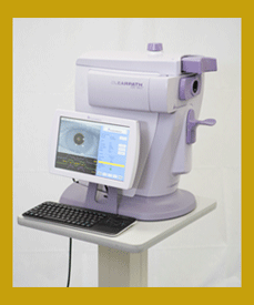
Integrate ClearPath DS-120 Biomicroscope Into Your Medical Model Practice
| Booth #534 |
"In my time with the ClearPath, I am seeing a high correlation between high AGEs in the lens, indicating multiple hyperglycemic events, and background diabetic hemorrhages. These results lead me to believe that this may be the first instrument that can help us delay the onset of diabetic retinopathy."
–John McCall, OD
The ClearPath DS-120 biomicroscope measures autofluorescence of the crystalline lens of the eye. In independent scientific studies published in peer-reviewed journals, elevated autofluorescence measurements have been linked to high levels of advanced glycosylated end products, which accumulate in the presence of diabetes and uncontrolled glucose over time.
By performing a science-based risk assessment for diabetes, the eye care professional is not strictly making a diagnosis but is providing invaluable information that may influence what lab tests are ordered by the primary care provider. Eye care providers, on average, see almost two times the number of patients a primary care provider sees in one year, making the eye care provider a key player in the front lines of diabetes detection. By detecting early and co-managing diabetes with a primary care provider, eye care providers will be able to recall the patient twice annually under American Optometric Association guidelines and receive reimbursements for quality of care.
In addition, primary care providers may benefit from good relationships with eye care providers on reimbursement. Under the Healthcare Effectiveness Data Information Set (HEDIS), set forth by the National Committee for Quality Assurance (NCQA), a physician's quality of care is based on the results of an overall 21-point audit of his or her care. Based on the results of the audit, a PCP can be paid an additional $25,000 to $50,000 per year, depending on the size of the practice. However, one quality measure in HEDIS—the number one area for which PCPs are routinely penalized—is failure to demonstrate that their patients with diabetes have received a dilated eye examination within the preceding 12 months. This failure is usually due to one or both of these reasons:
- No referral letter was sent by the PCP and/or eye doctor
- The patient never showed up for the eye examination
See the ClearPath demonstration at the booth (#534) or visit the Freedom Meditech website for more information.
New Technology: Sonomed Escalon
Breaking News: VuPad™ 510(k) Cleared by the FDA
| Booth #315 |
Sonomed Escalon will be taking orders on the new VuPad™ system at The Exchange. The system received its FDA approval earlier today.
"If a patient has a nuclear sclerotic cataract that is expanding, and the lens is expanding and closing off the angle, the patient has to be watched very closely. As the angle closes, the pressure shoots up, and the patient will fall into phacomorphic glaucoma. It will go away once the cataract is removed, but he or she needs to be monitored closely until that happens. And UBM is a great way to do that."
–John A. McCall, Jr., OD, "UBM Boosts Quality of Care for Glaucoma Practices",
Primary Care Optometry News, August 2013
Sonomed Escalon has been the leader in ophthalmic ultrasound for over 30 years. Its systems set the standards by which all others are measured. The new VuPad is no exception. This revolutionary portable device delivers exceptional image quality in a wide range of applications. It's also remarkably easy to use, thanks to intuitively designed touch screen controls. With VuPad, you can bring advanced ultrasound to more places—and more patients—than ever before.
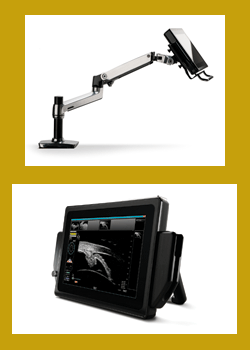
Industry-leading image quality: The better the image, the more accurate the diagnosis. VuPad combines Sonomed Escalon's superior UBM and newly enhanced B-scan image quality with an ultra high-resolution screen that has 25 percent larger viewing area than other portable ultrasound devices. Enhanced Focus Rendering™ allows you to capture both crisp still images and record video which can be carefully reviewed frame-by-frame.
An exceptional user experience: Concentrate on your patients, not on the controls. With an intuitive graphic interface, VuPad makes ultrasound simple and straightforward. The multi-touch screen puts important functions easily at your fingertips. You can also take advantage of innovative, smartphone-inspired features, like pinch zoom. VuPad also includes time-saving pre-set scan settings to automatically optimize image quality depending upon area of viewing interest.
Portable, flexible and adaptable: In ophthalmic practices, there's no such thing as an "ordinary day." The compact, ergonomic VuPad is designed to adapt. You can use it on tabletops with its back stand, or attach to carts or reticulating arms with the VESA mount. You choose the modalities you need and want – UBM, B-Scan, and/or A-Scan. You get the choice of 35 or 50 MHz Transducers (UBM), 12 or 20 MHz Probes (B-Scan) and Immersion or Soft-Touch Probes (for A-Scan). Dual-Band WIFI, Ethernet, USB, and Bluetooth allow you connect to other devices or your network. There's also plenty of room onboard to store images, with a hard drive that's 60 percent larger than other portable ultrasound devices.
Stop by the Sonomed Escalon booth (#315) at The Exchange.
Your Feedback Counts

Please take a moment to answer this two-question survey—even if you've done so before. It provides us the feedback to improve The Gazette.
©Vision Source L.P. 2014. All Rights Reserved.