
| Quarterly New Technology Update | March 27, 2015 |
In This Edition
- Special Tech Issue
- New Technology: Rabin Cone Test
- Product Review: XFraction
- Product Review: RHA™ Multi-Spectral Imaging Digital Ophthalmoscope
- Product Review: Cliradex™
- New Technology: Volk Eye Check
- Give Us Your Feedback
Quarterly New Technology Update
The Gazette
Archives


Contact Vision Source® at 888-558-2020 or [email protected]
Special Tech Issue
Read What's New and Impressive in the Quarterly New Technology Update
It is amazing the number of new technologies that we are able to see, as we travel to the various meetings across the country. Most of these companies with new technologies are eager to become vendor partners and offer these products and technologies to Vision Source® members. As a service to members, we will provide a quarterly update to members on the offerings from existing and new vendors with new technologies.
These products and services will be evaluated in a clinical setting, and we will try and point out the applications and advantages to these technologies so that you can gauge for yourself whether it is the right fit for your practice. All five of the companies whose products are featured in this issue will be exhibiting at The Exchange®, so make plans to drop by their booths and see the products and technology first-hand.
 |
 |
John A. McCall, OD |
Carl Spear, OD, FAAO |
New Technology: Rabin Cone Test
Innova Systems' Rabin Cone Test – Revolutionary Eye Disease Management
Innova Systems’ Rabin Cone Test is a revolutionary, Medicare-reimbursable technology for eye disease management using cone function.
Due to the sensitivity of the test, doctors can, for the first time, detect cone deficiencies present in eye disease, including macular degeneration, glaucoma and diabetic retinopathy, potentially before permanent damage occurs. With regular testing, changes in cone function can be used to monitor disease advancement and treatment efficacy.
The Rabin Cone Test can increase Medicare billings for color vision as well as drive further utilization of billable, structural tests by earlier detection. Practice-building is a natural result as patients look for preventative care with quantitative functional results.
The Rabin Cone Test was co-developed by Innova Systems and the US Air Force and has been researched extensively in clinical trials for AMD, glaucoma and diabetic retinopathy by Duke University, Shiga University, the US Army and others.

John McCall Jr., OD, has been evaluating Innova’s Rabin Cone Test in his practice for several months.

"This test has been utilized in my clinic for following macular degeneration, diabetic edema and glaucoma. The diabetes and AMD is straight forward. Distortion of the cones will reduce color recognition in both these diseases. Improvement will re-establish the color recognition, so we are able to monitor progression, as well as recovery. There also seems to be a correlation between GCC fallout and Color VA being compromised. There are lots of studies being done that validate this.
"The best part is the ROI: the cost of the Rabin Cone Test for Vision Source® is around $5,000 and the reimbursement average is $56. So for 1/10th the cost of an OCT, you can get $16 more than the OCT pays. This test gives valuable function information that complements the OCT."
Innova Systems will be showing the Rabin Cone Test at The Exchange® Meeting in Phoenix. For more information, please contact Innova Systems at 630-920-8880 or visit its website at innovasystemsusa.com.
Product Review: XFraction
XFraction – the True Wavefront Optimized Refraction by Marco
By John McCall Jr., OD
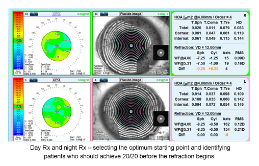
Marco is now the first to bring wavefront diagnostic technology into the automated refraction process and provide you with the True Wavefront Optimized Refraction, known as XFraction. XFraction is the process in which the full assessment of the total visual system is analyzed, mapped out and downloaded automatically into the phoropter for the final patient verification, which can be refined to the 0.12D. Now more than 70 percent of your patients will be shown the best Rx for either day or night, before you even say "1 or 2." The other 30 percent will now understand and SEE what is happening in the visual system and be able to achieve the patient the best possible RX, as well as the best recommendations for their vision. This new level of efficiency and accuracy as well as the low-order and high-order aberrations given to you from both the cornea and lens will tell you who is sitting in your exam chair before you even say "hello."
What is just as important as buying new technology is buying it from a company that can be your partner now and in the future. Marco has the team with the knowledge, experience and resources to not only bring this revolutionary technology to you but also be able to teach the information to you and your staff, support you and your practice and ensure that your technology integrates with your EMR now and in the future. Technology is constantly changing and it is important to be partnered with a company that invests and grows with you long term.
During The Exchange®, come and experience an XFraction exam and let us tell you about your vision! Or call 800-874-5274 to arrange for one of our technology experts to give you a consult.
Product Review: RHA™ Multi-Spectral Imaging Digital Ophthalmoscope
Advantages of Long Wavelength Fundus Autofluorescence

By Carl Spear, OD, FAAO
Annidis Corporation is launching a significant upgrade to the RHA™ Multi-Spectral Imaging Digital Ophthalmoscope with the addition of long wavelength fundus autofluorescence (FAF) imaging. Vision Source® members who currently have the RHA™ in their practices will be receiving software upgrades and training to enable this new capability in their clinics.
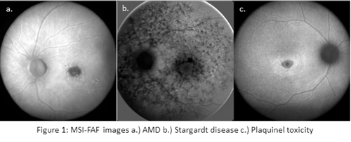
FAF imaging is used to document the presence of fluorophores such as lipofuscin in the eye. It provides physiological as well as functional information regarding the health of the retina, primarily the retinal pigment epithelium (RPE) and the photoreceptors. The prognosis of some diseases such as AMD, Stargardt disease and Plaquinel toxicity (Figure 1), as well as multiple other retinal dystrophies and degenerations, can be determined by the presence or absence of autofluorescence.
 Visible light may be used to fluoresce lipofuscin, the byproduct of RPE outer segment phagocytosis1 at various visible wavelengths, but there are some limitations at shorter wavelengths. The RHA™ uses an absorption and re-emission process in which the retina is illuminated with an average wavelength of 600 nm. A red barrier filter blocks the strong signal from the retina in this wavelength region of the spectrum and passes the light back in the 660 nm region. This improves the signal to noise ratio and hence the image quality.2
Short wavelength FAF is attenuated for both excitation and emission due to scatter from the crystalline lens.2 With cataracts, particularly nuclear opacities3, scatter is even more pronounced. Moreover, the lens itself contributes to the overall ocular autofluorescence, not just fundus autofluorescence, when short wavelengths are used.
Additionally, the FAF signal is reduced in the macular region with short wavelength FAF due to the absorption of blue light by the macular pigments lutein and zeaxanthin in the neurosensory retina.4 Another limitation is the interference of fluorescein angiography on short wavelength FAF, because the excitation wavelength may be within the absorption spectrum of the dye.5 Long wavelength FAF imaging can penetrate through the crystalline lens and macular pigment to reveal hyper- and hypoautofluorescent lesions and is not subject to interference from fluorescein dye.
The primary fluorescent component of lipofuscin is A2E [N-retinylidene-N-retinylethanolamine]).1 The excitation and emission spectral wavelengths of A2E are longer than that of the other lipofuscin fluorophores, making it preferentially selected for long wavelength excitation.6 This dominance at long wavelengths makes it the ideal target for MSI-FAF imaging, allowing for definitive fluorophore discrimination.7 Long wavelength FAF of A2E in the deep retina improves the contrast of the retinal vasculature over short wavelength FAF.7
For more information, visit annidis.com or email [email protected] or call 1-855-ANNIDIS (266-4347) ext. 800.
References:
1. Weiter, JJ et al. Retinal Pigment Epithelial Lipofuscin and Melanin and Choroidal Melanin in Human Eyes. Invest Ophthalmol Vis Sci. 1986; 27: 145-152.
2. Holz, FG et al. (2007) Atlas of Fundus Autofluorescence Imaging. Springer-Verlag Berlin Heidelberg.
3. Ruiz-Moreno JM et al. Fundus autofluorscence in age-related macular degeneration. www.amdbook.org
4. Kellner, U, Kellner, S & Weinitz, S. Fundus autofluorescence (488nm) and near infra-red autofluorescence (787nm) visualize different retinal pigment epithelium alterations in patients with age-related macular degeneration. Retina. 2010; 30(1): 6-15.
5. Optimized Filters Move Autofluorescence Imaging Forward. Retinal Physician. October 2009.
6. Sparrow, JR & Duncker, T. Fundus Autofluorescence and RPE Lipofuscin in Age-Related macular Degeneration. J.Clin. Med. 2014; 3(4): 1302-1321.
7. Schweitzer, D et al. Towards Metabolic Mapping of the Human Retina. Experimental Ophthalmology, University of Jena, Bachstr. 18, Germany.
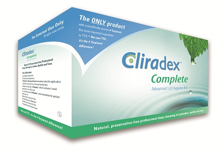
Product Review: Cliradex™
Innovative Protocol for Lid Margin Disease
By Carl Spear, OD, FAAO
Bio-Tissue’s Cliradex® Complete Advanced Lid Hygiene Kit is the newest addition to its Cliradex® product line, the only commercially available products that isolates 4-Terpineol, the most important ingredient in tea tree oil scientifically proven to help manage symptoms of lid margin diseases (LMD), including blepharitis, demodex, rosacea and dry eye.1
In the United States alone, there are approximately 80 million people with blepharitis, many of whom are elderly. According to literature, between 40 percent and 80 percent of these cases are caused by demodex, which, due to the fact that it’s microscopic and the mites can’t be seen by the naked eye, is largely underdiagnosed, leaving patients in much discomfort. In more severe chronic cases, patients present with symptoms such as redness, swelling and itching that are hard to manage and require immediate attention.
The Cliradex Complete Advanced Lid Hygiene Kit is a professional, comprehensive lid hygiene protocol featuring a formulation gel called Cliradex® Advanced Care that includes a stronger concentration of 4-Terpineol for in-office application by a doctor or trained technician. It is used for moderate to severe cases of lid margin diseases and also contains ingredients for easy removal of lid margin debris and make up. The kit comes with a dual-sided applicator, doctor and patient materials and a carton of Cliradex® lid wipes for patients to use at home for ongoing management of their symptoms.
“This innovative in-office treatment helps to effectively manage symptoms of moderate to severe cases of lid margin disease, leading to better quality of life for my patients,” said Scott Schachter, OD, of Advanced Eye Care, a Vision Source® member practice in Pismo Beach, California. “To increase patient compliance, I make a strong recommendation, thoroughly explain the treatment protocol and why it’s important, and then ask the patient if they have questions. This approach leads to greater patient adoption, faster healing and improved ocular health.”
Tea Tree Oil and the 4-Terpineol Difference
Although tea tree oil has been widely used in eye care practices, it can burn and be toxic to the cornea if the concentration isn’t reduced. Studies have shown that 4-Terpineol, one of major components of tea tree oil, has the highest impact at the lowest concentration. Unlike traditional lid care products with no active ingredient, 4-Terpineol, the active ingredient in the Cliradex® line of products, provides a therapeutic response with high success rates. This is because the ingredients in tea tree oil neutralize each other, rendering them less effective. Isolated 4-Terpineol, on the other hand, is scientifically proven to manage symptoms of lid margin diseases.
Cliradex products are natural, preservative-free lid, lash and facial cleansers; more information can be found at www.cliradex.com. To learn more about the Vision Source® special introductory pricing and to receive a reimbursement guide for Cliradex Complete Advanced Lid Hygiene Kit, request a free trial, and schedule a sales representative visit, call Dion Maclin at 888-296-8858.
References
1 Tighe Sean, et al, Terpinen-4-ol is the Most Active Ingredient of Tea Tree Oil to Kill Demodex Mites, Translational Vision Science and Technology, August 2013, Vol. 2, No. 7
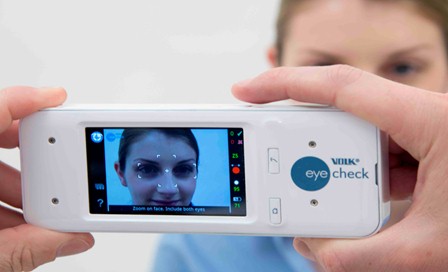
New Technology: Volk Eye Check
The Volk Eye Check: How data acquisition can benefit your patient work flow
by John McCall Jr., OD
The Volk Eye Check is an easy-to-use, handheld diagnostic assistance device that provides optometrists with automatic real-time measurements of seven key eye features – including pupil size, visible iris diameters, eye apertures, lid positions and manifest strabismus -- to assist in detecting and diagnosing various eye conditions as well as improving the efficiency of contact lens fitting.
Pediatric Screening
Performing the cover test on our newest and youngest patients can be a challenging experience. The Volk Eye Check is suitable for pediatric applications down to 6 months with the ability to indicate conditions such as strabismus, ptosis and anisocoria.
Contact Lens Fitting
It is often too late that we find out about a patient’s discomfort with a contact lens prescription. Away from the practice, there is little that can prevent a patient from dropping out of repeat prescriptions, as around 20 percent of our patients do.
New research from leading schools is showing that increased data capture can ensure a new patient has the best possible fit on a first visit. HVID, pupil size and eccentricity, lid position and sagittal depth can all play a role in a well-fitted contact lens.
Pupil size is very difficult to measure, sometimes simply because the patient has dark iridies and measurement with a millimeter ruler is inaccurate. The Volk Eye Check accurately measures the pupil size to within +/-0.20mm.
The Volk Eye Check provides vertical, oblique and horizontal visible iris diameter which is a key parameter for good fitting of contact lenses, especially multifocal lenses. There is an option when using the Contact Lens module to pre-input keratometry readings and automatically generate the corneal sag for each eye. The Volk Eye Check improves the quality of care and saves chair time in the busy office.
Contact Lens Case History
54-year-old male new patient. Low hypermetrope with minimal astigmatism. Previously unsuccessfully fitted elsewhere with monovision and multifocal contact lenses. He was generally unhappy with his vision at all distances and wanted a second opinion to be fitted with multifocal contact lenses.
Volk Eye Check revealed the reason for failure. The patient had large HVIDs with resulting unusually high sag, leading to poor centration, combined with small pupils in normal lighting conditions. Failure should have been expected.
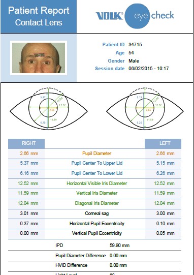
This combination of data provided clinical evidence that that this patient would be unsuitable for conventional multifocals. Rather than waste chair-time trying ‘off the shelf’ lenses the patient was advised that custom lenses would be the most appropriate option.
Summary of the clinical power of the device
• Quickly identifies areas for investigation (strabismus, anisocoria, ptosis) and provides objective electronic record of the readings
• Assists the clinician in identifying best-fit contact lenses; reduces dropout rates and saves chair-time and money; assists in the design of custom made lenses
• Objective EMR of eye features and eye conditions for compliance, referrals and litigation
Suitable for babies and young children – even at 6 months old
• Monitoring of treatment progress (pre-op/post-op)
• Reduces inter-clinician variability – standardize testing across practices and clinicians in optometry chains
• Oculoplastics: accurate measurement of lid position for lid and cosmetic surgery.
For more information about Volk Eye Check, visit www.volk.com/eyecheck, phone Volk direct at 1-800-345-8655 (+1 440-942-6161 outside the United States), or contact your Authorized Volk Distributor.
Your Feedback Counts

Please take a moment to answer this two-question survey—even if you've done so before. It provides us the feedback to improve The Gazette.
©Vision Source L.P. 2015. All Rights Reserved.